The security strength of an improved optical cryptosystem
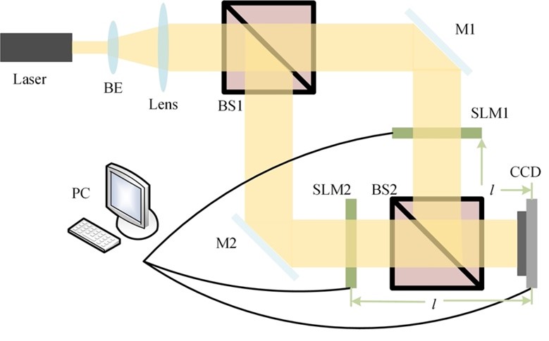 In the last few years, due to the enormous development in communication technology, the sharing, and transmission of information have increased immensely. The information can be transferred in various forms, such as text, audio, video, and images. Mostly, the information or data is transmitted through open channels, which increases the possibility of illegal interception, fabrication, and modification of the original information. Thus, to avoid unauthorised access or alteration of data, the development of secure transmission systems is very important.
The latest research from the Department of Physics evaluates the security strength of an improved optical cryptosystem based on interference. Assistant Prof Dr Ravi Kumar has published a paper, Security analysis on an interference-based optical image encryption scheme, in the Applied Optics journal, with an impact factor of 1.905.
Dr Ravi Kumar’s research is focused on the area of optical information processing and optical metrology. He studies and designs new optical cryptosystems with enhanced security features. For that, he uses various optical aspects and techniques, such as interference, diffractive imaging, polarization, computational imaging, etc. Alongside this, he also works in the area of digital holography and incoherent imaging. In this, he designs and develops new optical systems for imaging applications, such as super-resolution imaging, biomedical imaging, 3D imaging, telescopic applications, object detection, reconstruction, etc.
Explanation of the Research
Optical systems have been studied extensively for image encryption and found to be more reliable and efficient than their digital counterparts, such as parallel processing, capable of processing 2D data, multi-parameters capabilities (i.e., phase, wavelength, polarization, etc.), and can be employed as the security keys. The usage of biometric authentication in daily life, credit cards, fingerprint authentication, email/bank passwords, etc.; all need to be secured. This research can play an important role in designing a sophisticated cryptosystem for future technologies. Moreover, another direction of the research i.e., optical imaging, can be translated to design new low-cost biomedical devices (endoscopes, microscopes, biomedical sensors, etc.) which can have a significant social impact.
In the future, Dr Ravi Kumar will be focusing on the development of a new robust optical cryptosystem and designing new attack algorithms for existing optical encryption techniques. Additionally, he is also designing new optical imaging systems with better signal-to-noise ratios and improved resolution.
Abstract
In this paper, the security strength of an improved optical cryptosystem based on interference has been evaluated. The plaintext was encoded into a phase-only mask (POM) and an amplitude mask (AM). Since the information of the plaintext cannot be recovered directly when one of the masks is released in the decryption process of an improved cryptosystem, it seems that it is free from the silhouette problem. However, researchers found that the random phase mask (RPM) that served as the encryption key is not related to the plaintext; thus, it is possible to recover the RPM firstly using the known-plaintext attack (KPA). Moreover, the POM and the AM generated in the encryption path only contains the phase and amplitude information, respectively; thus, these can be utilised as additional constraints in the proposed iterative process. Based on these findings, researchers have demonstrated two new kinds of hybrid attacks to crack the cryptosystem, i.e., a KPA and an iterative process with different constraints. To the best of our knowledge, it was the first time that the existence of a silhouette problem in the cryptosystem under study had been reported. Researchers have validated their attacks through numerical simulation.
Collaborations
Dr Xiong Yi, Jiangnan University, Wuxi 214122, China
- Published in Departmental News, News, Physics News, Research News
‘The modern Kautilya of India’: Dr C Rangarajan on India’s economic development
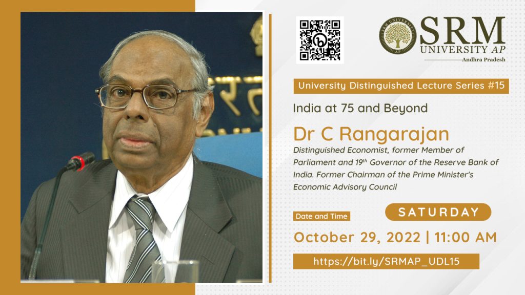 The fifteenth edition of University Distinguished Lecture series on the topic “India at 75 and beyond”, was held on October 29, 2022 to celebrate the magnificent growth displayed by India. The session was addressed by Dr C Rangarajan, renowned economist and former Governor of Reserve Bank of India. The intense and inspiring lecture highlighted the importance of reflection on the past and articulation of our vision for our future to enable rapid progression on economic development.
Dr C Rangarajan gave a comprehensive outlook on the economic performance of India since independence. “India has made momentous progress on reducing multidimensional poverty. The incidents of multidimensional poverty were almost reduced by half to almost 27.5% during 2005-06 and 2015-16 period due to deeper progress among the poorest. Thus within 10 years, the number of poor people in India fell by more than 270 million, a truly massive achievement,” he stated during the lecture.
Dr Rangarajan further expounded on the importance of reform agendas and measures, the subsisting triad of economic policies and the future challenges of progressing into being a developed nation. The lecture was followed by a Q & A session moderated by Dr S Ananda Rao and Dr Erra Kamal Sai Sadharma from the Department Economics.
Prof Kamaiah Bandi, Dean-School of Liberal Arts and Social Sciences applauded Dr Rangarajan on being a unique distinction of shaping and motivating five generations of intellectual cohort. “Dr C Rangarajan has successfully brought down the gap between theory and practice in his capacity as Governor of RBI and various other important positions he has held for our nation. We as SRM AP look forward to your remarkable experience and knowledge to incubate motivation in our students.”
SRM University-AP has actively promoted a cumulative intellectual ecosystem and interdisciplinary education. “The principal objective of the University Distinguished Lecture series is to impel research scholars, students from all around the world to undertake progressive measures for the holistic development of our nation”, said Honourable Vice Chancellor, Prof Manoj K Arora in his welcome address.
Prof D Narayana Rao, Pro-Vice-Chancellor, SRM University-AP concluded the event by addressing Dr C Rangarajan as ‘the modern Kautilya of India’ and presented a memento on behalf of the institution as a token of respect and appreciation for his esteemed presence at the fifteenth edition of the University Distinguished Lecture series.
Doctoral scholar secures visiting fellowship
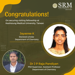
Exposure to international research opportunities promotes empirical learning at an impeccable level. International research ventures aid scholars to explore novel research avenues enabling a transformative progress for society through the field of science. The Department of Chemistry is glad to announce that Ms Jayasree K, PhD scholar, has been accepted for Short-Term Research Internship (STRI) for a period of six months from the Research Center of Environmental Medicine, Kaohsiung Medical University, Taiwan.
Ms Jayasree has been elevated in receiving the offer and delightfully keen on the new avenues she could explore through this opportunity. She is currently working in the field of surface-enhanced Raman spectroscopy (SERS). In this particular research area, her major research objective is the design and development of a novel SERS substrate for food and bioanalysis.
“My internship mentor, Prof. Vinoth Kumar, KMU University is an expert in mass spectroscopy and High-performance liquid chromatography (HPLC). Therefore, I have an option to hyphenate the Raman technique along with mass spectroscopy which leads Raman research to the next level for various applications”, commented Ms Jayasree on this incredible opportunity.
Her internship at Kaohsiung Medical University (KMU) is based on the motive of research on food and environmental toxicity which would provide guidance on her first research project in the field of food analysis.
She has offered her sincere gratitude to her supervisor, Dr Rajapandiyan JP, Department of Chemistry for his constant support and advice from the application process to proposal writing, experimental planning etc. She also thanked SRM University- AP in providing support through the process and extending travel allowance and guidance.
Ms Jayasree utilizes this great opportunity to explore and discover herself, developing both personally and professionally. Through this internship she hopes to learn new skills, expand her knowledge in the field of research and explore career options in Taiwan.
- Published in Chemistry-news, Departmental News, News, Students Achievements
Classification of brain tumours using fine tuned ensemble of ViTs
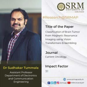
Primary brain tumours make up less than 2% of cancers and statistically occur in around 250,000 people a year globally. Medical resonance imaging (MRI) plays a pivotal role in the diagnosis of brain tumours and advanced imaging techniques can precisely detect brain tumours. On this note, Dr Sudhakar Tummala, Assistant Professor, Department of Electronics and Computer Engineering, has published a paper titled, “Classification of Brain Tumour from Magnetic Resonance Imaging using Vision Transformers Ensembling” in the journal Current Oncology having an impact factor of 3.1. The paper highlights the pioneering breakthrough made in the development of vision transformers (ViT) in enhancing MRI for efficient classification of brain tumours, thus reducing the burden on radiologists.
Abstract of the paper
The automated classification of brain tumours plays an important role in supporting radiologists in decision making. Recently, vision transformer (ViT)-based deep neural network architectures have gained attention in the computer vision research domain owing to the tremendous success of transformer models in natural language processing. Hence, in this study, the ability of an ensemble of standard ViT models for the diagnosis of brain tumours from T1-weighted (T1w) magnetic resonance imaging (MRI) is investigated. Pretrained and fine tuned ViT models (B/16, B/32, L/16, and L/32) on ImageNet were adopted for the classification task. A brain tumour dataset from figshare, consisting of 3064 T1w contrast-enhanced (CE) MRI slices with meningiomas, gliomas, and pituitary tumours, was used for the cross-validation and testing of the ensemble ViT model’s ability to perform a three-class classification task. The best individual model was L/32, with an overall test accuracy of 98.2% at 384 × 384 resolution. The ensemble of all four ViT models demonstrated an overall testing accuracy of 98.7% at the same resolution, outperforming individual model’s ability at both resolutions and their ensemble at 224 × 224 resolution. In conclusion, an ensemble of ViT models could be deployed for the computer-aided diagnosis of brain tumours based on T1w CE MRI, leading to radiologist relief.
A brief summary of the research in layperson’s terms
Brain tumours (BTs) are characterised by the abnormal growth of neural and glial cells. BTs causes several medical conditions, including the loss of sensation, hearing and vision problems, headaches, nausea, and seizures. There exist several types of brain tumours, and the most prevalent cases include meningiomas (originate from the membrane surrounding the brain), which are non-cancerous; gliomas (start from glial cells and the spinal cord); and glioblastomas (grow from the brain), which are cancerous. Sometimes, cancer can spread from other parts of the body, which is called brain metastasis. A pituitary tumour is another type of brain tumour that develops in the pituitary gland in the brain, and this gland primarily regulates other glands in the body. Magnetic resonance imaging (MRI) is a versatile imaging method that enables one to noninvasively visualise inside the body, and is in extensive use in the field of neuroimaging.
There exist several structural MRI protocols to visualise inside the brain, but the prime modalities include T1-weighted (T1w), T2-weighted, and T1w contrast-enhanced (CE) MRI. BTs appear with altered pixel intensity contrasts in structural MRI images compared with neighbouring normal tissues, enabling clinical radiologists to diagnose them. Several previous studies have attempted to automatically classify brain tumours using MRI images, starting with traditional machine learning classifiers, such as support vector machines (SVMs), k-nearest-neighbour (kNN), and Random Forest, from hand-crafted features of MRI slices. With the rise of convolutional neural network (CNN) deep learning model architectures since 2012, in addition to emerging advanced computational resources, such as GPUs and TPUs, during the past decade, several methods have been proposed for the classification of brain tumours based on the finetuning of the existing state-of-the-art CNN models, such as AlexNet, VGG16, ResNets, Inception, DenseNets, and Xception, which had already been found to be successful for various computer vision tasks.
Despite the tremendous success of CNNs, they generally have inductive biases, i.e., the translation equivariance of the local receptive field. Due to these inductive biases, CNN models have issues when learning long-range information; moreover, data augmentation is generally required for CNNs to improve their performance due to their dependency on local pixel variations during learning.Therefore, in this work, the ability of pretrained and fine tuned ViT models, both individually and in an ensemble manner, is evaluated for the classification of meningiomas, gliomas, and pituitary tumours from T1w CE MRI at both 224 × 224 and 384 × 384 image resolutions.
Dr Sudhakar Tummala has mentioned the social implications of the research by expounding that the computer-aided diagnosis of brain tumours from T1w CE MRI using an ensemble of fine tuned ViT models can be an alternative to manual diagnoses, thereby reducing the burden on clinical radiologists. He also explains the future prospects of his research, which is to add explainability to the ensemble model predictions and to develop methods for precise contouring of tumour boundaries.
Details of Collaborations
Prof Seifedine Kadry, Department of Applied Data Science, Noroff University College, Kristiansand, Norway.
Dr Syed Ahmad Chan Bukhari, Division of Computer Science, Mathematics and Science, Collins College of Professional Studies, St. John’s University, New York, USA.
- Published in Departmental News, ECE NEWS, News, Research News
Novel antenna-duplexer for off-body communication
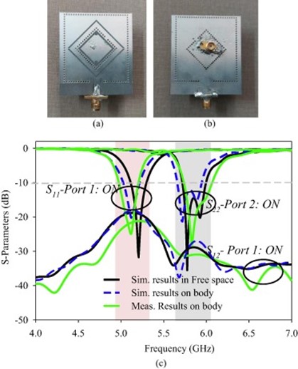
With the recent advancements in modern wireless body area network (WBAN) communication, the demand for compact low-profile wireless computing devices has witnessed a vast increase. Consequently, the antennas which play a critical role in this network are developed with different polarization in distinct frequency bands so as to maintain better reliability of communication links. Dr Divya Chaturvedi, Assistant Professor, Department of Electronics and Communication Engineering, has published a paper titled, “A Dual-Band Dual-Polarized SIW Cavity-Backed Antenna-Duplexer for Off-body Communication” as first author in the Q1 Journal AEJ – Alexandria Engineering Journal having an impact factor of 6.77. The paper discusses the self-duplexing antennas, offering two channels for concurrent transmission and reception, leading to a simple and compact transceiver.
Abstract
A novel dual-band, dual-polarized antenna-duplexer scheme is intended to be used for WLAN 802.11a and ISM band applications using Substrate Integrated Waveguide (SIW) Technology. The antenna consists of two planar SIW cavities of different dimensions where a smaller sized diamond- shaped cavity is inserted inside the larger rectangular cavity to share the common aperture area. The diamond-ring shaped slots are etched in each cavity for radiation. The larger diamond ring slot is excited with a microstrip feedline to operate at 5.2 GHz while the smaller slot is excited with a coaxial probe to operate at 5.8 GHz. The antenna produces linear polarization at 5.2 GHz (5.1–5.3 GHz) due to the merging of TE 110 and TE 120 cavity modes while circular polarization around 5.8 GHz due to orthogonally excited TM100 and TM010 modes (5.68–5.95 GHz). The slots are excited in an orthogonal fashion to maintain a better decoupling between the ports (i.e. –23 dB). The performance of the antenna has been verified in free space as well as in the vicinity of the human body. The antenna offers the gain of 6.2 dBi /6.6 dBi in free space and 5.8 dBi / 6.4 dBi on-body at lower-/ higher frequency-bands, respectively. Also, the specific absorption rate (SAR) is obtained < 0.245 W/Kg for 0.5 W input power averaged over 10 mW/g mass of the tissue. The proposed design is a low-profile, compact single-layered design, which is a suitable option for off-body communication.
Explanation of the research in layperson’s terms
- This antenna can operate in dual radio frequency bands at 5.2 GHz and 5.8 GHz respectively.
- The antenna can be used in the medical instrument to make it wire-free.
- The antenna is compact in size, thus can be accommodated in a small space.
- The antenna can operate simultaneously at both the frequency bands, thus at the same time it can help in forming links with another on-body antenna and makes the link with Wi-Fi.
- The antenna is validated in terms of Specific absorption rate, hence it is safe to use on the human body.
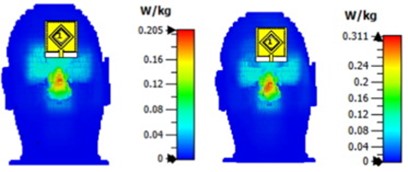
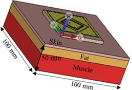
The paper further expounds on the social implication of this innovative research. Dr Chaturvedi explains that the antenna, being dual-band and dual-polarized, can function as a transceiver circuit. Due to different polarization, it can operate in both the frequency bands simultaneously without affecting the performance. In the first frequency band at 5.2 GHz, it can link with Wi-Fi and in the second frequency band at 5.8 GHz, it is able to communicate with antennas placed in other medical instruments which are used in the vicinity of the human body.
Collaborations
1. Dr Arvind Kumar, Assis. Professor, b Department of Electronics and Communication
Engineering, VNIT Nagpur, India
2. Dr Ayman A Althuwayb, Department of Electrical Engineering, College of Engineering,
Jouf University, Sakaka, Aljouf 72388, Saudi Arabia
- Published in Departmental News, ECE NEWS, News, Research News
Bioinspired GO/Au nanocomposite synthesis
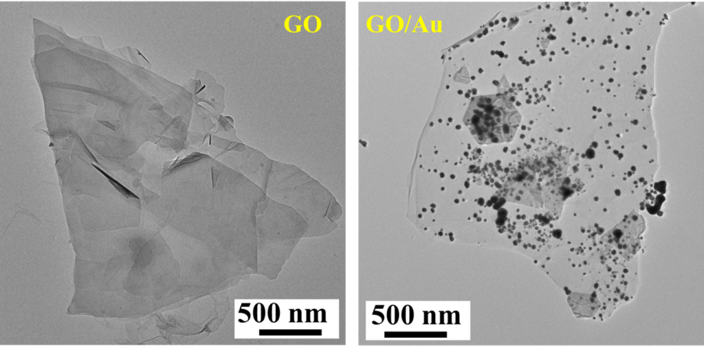
Nanocomposites are the heterogeneous materials that are produced by the mixtures of polymers with inorganic solids that are multi-phased with two or three dimensions of less than 100 nanometers (nm). Nanocomposites offer advanced technologies in enhancing several industrial sectors like automobile, construction, electronics and electrical, food packaging, and technology transfer, yet its sustainable and environment-friendly nature provides a great deal for mankind. Dr Imran Uddin, Post Doctoral fellow, Department of Physics, has published a paper titled “Bioinspired GO/Au nanocomposite synthesis: Characteristics and use as a high-performance dielectric material in nanoelectronics” in the South African Journal of Botany, having an impact factor of 3.11. The paper demonstrated that GO-based materials are better constituents for nanocomposite synthesis and facilitate in enhancing the performance of electrical devices and energy storage systems.
Abstract
A bioinspired method was used to synthesise a graphene oxide (GO) based noble metal (Au) nanocomposite (GO/Au nanocomposite) using chemically exfoliated graphene oxide as the base matrix and gold (Au) nanoparticles. GO’s structural properties and morphology and the GO/Au nanocomposite were determined using XRD, TEM, SEM, EDAX, FTIR, and TGA analysis. LCR analysis was used to characterise the electrical characteristics of GO dielectric features as a function of frequency. The dielectric permittivity and electrical conductivity of GO were very frequency-driven. The results demonstrated that GO has direct current and Correlated Barrier Hopping conductivity processes in the low and high-frequency bands. The dielectric constant of the GO/Au nanocomposite shows that the bioinspired approach includes organic macromolecules capable of modest GO reduction and so modifying the C/O ratio, resulting in an enhancement in the matrix’s dielectric characteristics. This work shows that GO-based materials can be used to scale up high-performance electronic devices, as well as electrical and energy storage systems.
Explanation of the research in layperson’s terms
Energy consumption has increased multifold over the past few years. With increased consumption, the need for energy production and storage has become a pressing priority in the current generation. Dr Imran Uddin’s work aims to propose an idea to synthesise a mixture of two energy-storing materials (gold and carbon) at room temperature. Keeping in view the mentioned aim, he has used plant seeds to create this energy-storing mixture, also known as dielectric material in scientific terms. Through various analyses, he has noticed that this material is able to store electric energy at a lower frequency than the parent material. The superiority of this material comes into play in that when it expires, it can be easily disposed of without creating pollution, which goes hand in hand with the ultimate aim to develop sustainable energy-storing devices.
Dr Imran Uddin has mentioned the practical implication of the groundbreaking research. Capacitors are electronic devices that store electric energy in the form of charges. When a capacitor is linked to a charging circuit, it can store electric energy and release that stored energy when attached to an external circuit (like cars, fans, nuclear weapons, etc.), allowing it to be used as a temporary battery. Moreover, the synthetic GO/Au nanocomposite has the potential to be used as a capacitor material in biomedical applications (defibrillators, blood gas analyzers, pacemakers, biomedicines, etc.), as well as other fields where non-toxicity is essential.
The future prospects of Dr Imran Uddin’s research view an ambitious plan to manufacture more materials at room temperature using the green synthesis root. He also intends to investigate the electrochemical characteristics of environmentally benign materials in the field of electrochemical energy storage, such as supercapacitors and batteries.
Collaborations
University of Pannonia, Hungary
- Published in Departmental News, News, Physics News, Research News
Detecting Breast cancer subtypes using an innovatory ensemble of SwinTs
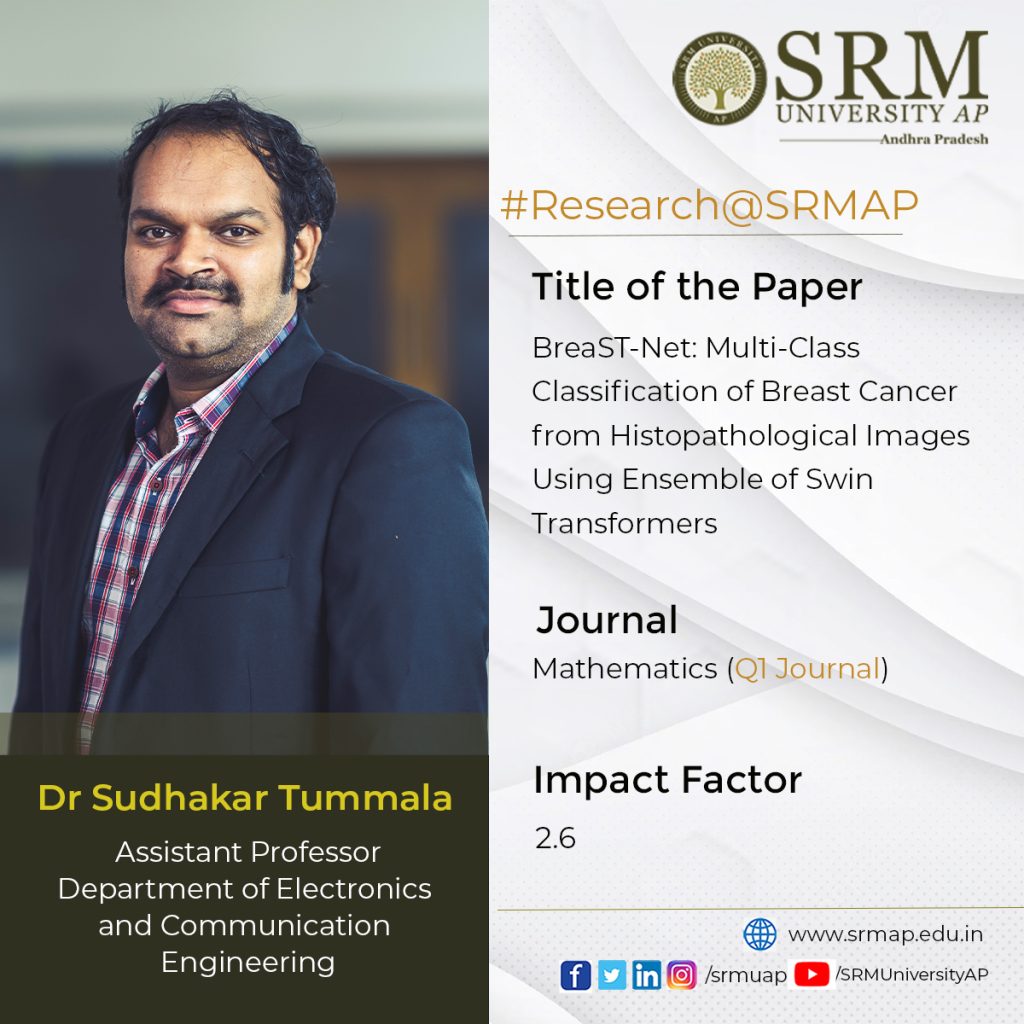
Breast cancer (BC) is one of the most common types of cancer among women with a high mortality rate. Histopathological analysis facilitates the detection and diagnosis of BC but is a highly time-consuming specialised task, dependent on the experience of the pathologists. Hence, there is a dire need for computer-assisted diagnosis (CAD) to relieve the workload on pathologists. Dr Sudhakar Tummala, Assistant Professor, Department of Electronics and Communication Engineering, has conducted breakthrough research on this domain in his paper titled BreaST-Net: Multi-Class Classification of Breast Cancer from Histopathological Images Using Ensemble of Swin Transformers published in the Q1 Journal Mathematics, having an Impact Factor of 2.6.
Abstract
Breast cancer (BC) is one of the deadly forms of cancer and a major cause of female mortality worldwide. The standard imaging procedures for screening BC involve mammography and ultrasonography. However, these imaging procedures cannot differentiate subtypes of benign and malignant cancers. Therefore, histopathology images could provide better sensitivity toward benign and malignant cancer subtypes. Recently, vision transformers are gaining attention in medical imaging due to their success in various computer vision tasks. Swin transformer (SwinT) is a variant of vision transformer that works on the concept of non-overlapping shifted windows and is a proven method for various vision detection tasks. Hence, in this study, we have investigated the ability of an ensemble of SwinTs for the 2- class classification of benign vs. malignant and 8-class classification of four benign and four malignant subtypes, using an openly available BreaKHis dataset containing 7909 histopathology images acquired at different zoom factors of 40×, 100×, 200× and 400×. The ensemble of SwinTs (including tiny, small, base, and large) demonstrated an average test accuracy of 96.0% for the 8-class and 99.6% for the 2-class classification, outperforming all the previous works. Hence, an ensemble of SwinTs could identify BC subtypes using histopathological images and may lead to pathologist relief.
A brief summary of the research in layperson’s terms
Breast cancer (BC) is the second deadliest cancer after lung cancer, causing morbidity and mortality worldwide in the women population. Its incidence may increase by more than 50% by the year 2030 in the United States. The non-invasive diagnostic procedures for BC involve a physical examination and imaging techniques such as mammography, ultrasonography and magnetic resonance imaging. However, the physical examination may not detect it early, and Imaging procedures offer low sensitivity for a more comprehensive assessment of cancerous regions and identification of cancer subtypes. Histopathological imaging via breast biopsy, even though minimally invasive, may provide accurate identification of the cancer subtype and precise localization of the lesion. However, this manual examination by the pathologist could be tiresome and prone to errors. Therefore, automated methods for BC subtype classification are warranted.
Deep learning has revolutionised many areas in the last decade, including healthcare for various tasks such as accurate disease diagnosis, prognosis, and robotic-assisted surgery. There were studies based on deep convolutional neural networks (CNN) for detecting BC using the aforementioned imaging procedures. However, CNNs exhibit inherent inductive bias and are variant to translation, rotation, and location of the object of interest in the image. Therefore, image augmentation is generally applied while training CNN models, although the data augmentation may not provide expected variations in the training set. Hence, self-attention based deep learning models that are more robust towards the orientation and location of an object of interest in the image are rapidly growing.
SwinTs are an improved version of earlier vision transformer (ViT) architecture and are hierarchical vision transformers using shifted windows that work based on self-attention. For efficient modelling, self-attention within local windows was proposed and computed, and to evenly partition the image, the windows are arranged in a non-overlapping manner. The window-based self-attention has linear complexity and is scalable. However, the modelling power of window-based self-attention is limited because it lacks connections across windows. Therefore, a shifted window partitioning approach that alternates between the partitioning configurations in consecutive Swin transformer blocks was proposed to allow cross-window connections while maintaining the efficient computation of non-overlapping windows. The shifted window scheme in Swin transformers offers increased efficiency by restricting self- attention computation to local windows that are non-overlapping while also facilitating a cross-window connection. Overall, the SwinT network’s performance was superior to that of the standard ViTs.
Therefore, the paper analyses the ability of an ensemble of Swin transformer models (BreaST-Net) for the automated multi-class classification of BC by investigating histopathological images. The work dealt with both benign and malignant subtypes. Further, the benign cancer subtypes include fibroadenoma, tubular adenoma, phyllodes tumour, and adenosis. Whereas the malignant subtypes contain ductal carcinoma, papillary carcinoma, lobular carcinoma, and mucinous carcinoma.
Social implications of the research
Dr Sudhaker Tummala explains that the computer-aided subtyping of breast cancer from histopathology images using an ensemble of fine-tuned SwinT models can be an alternative to manual diagnoses, thereby reducing the burden on clinical pathologists.
Collaborations
- Prof. Seifedine Kadry, Department of Applied Data Science, Noroff University College, Kristiansand, Norway
- Dr Jungeun Kim, Division of Computer Science, Department of Software, Kongju National University, Korea
In the future, Dr Tummala will advance his research to add explainability to the ensemble model predictions and also to develop models that can work on fewer data samples.
- Published in Departmental News, ECE NEWS, News, Research News
Dr Anil K Suresh and team exploring novel domains of research at SRM AP!
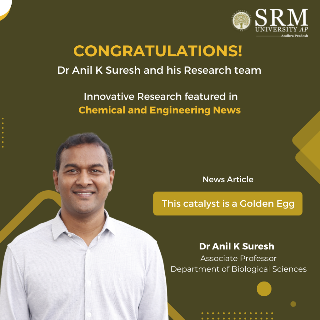
SRM AP proudly congratulates Dr Anil K Suresh, Associate Professor, Department of Biological Sciences and his cohort of research scholars for their rare achievement of having their paper featured in the prestigious weekly news magazine Chemical and Engineering News (ACS-C&EN). The news article titled “This catalyst is a Golden Egg“, edited by Prachi Patel highlights the innovative research conducted by Dr Anil K Suresh and his team on developing a low-cost, sustainable catalyst by infusing eggshells with gold nanoparticles that can be reused and eventually recycled.
The research paper titled Sustainable Bio-Engineering of Gold structured Wide-Area Supported Catalyst for Hand-Recyclable Ultra-Efficient Heterogeneous Catalysis (ACS Appl. Mater. Interfaces 2022, DOI: 10.1021/acsami.2c13564) highlights the team’s breakthrough advance in impregnating eggshells with gold nanoparticles to develop a cheap, and reusable ‘mega catalyst’. The research has used the robust “mega catalyst” to detoxify dye waste and run other organic reactions by dropping the eggshell catalyst into reaction solutions.
Dr Suresh and colleagues dipped waste eggshells collected from restaurants in a suspension containing gold chloride for 6 hours. Consequently, the amino acids on the shells reduced the gold ions to form crystalline gold nanoparticles. Dr Suresh attested this method to be one of the simplest syntheses of a supported catalyst that was ever reported. He emphasised that he and his colleagues have also grafted catalytic silver and copper oxide on eggshells and are now working on methods to do this for platinum and palladium.
SRM University-AP manifests pioneering advancement in innovative domains of research in the field of science and technology. The breakthrough research by Dr Anil K Suresh and his research team is of outstanding achievement and incredible addition to the institution’s laurels in the field of translational research.
- Published in Biology News, Departmental News, Faculty Achievements, News, Research News
EEE Students Visited Dr Narla Thatharao Thermal Power Station (NTTPS)
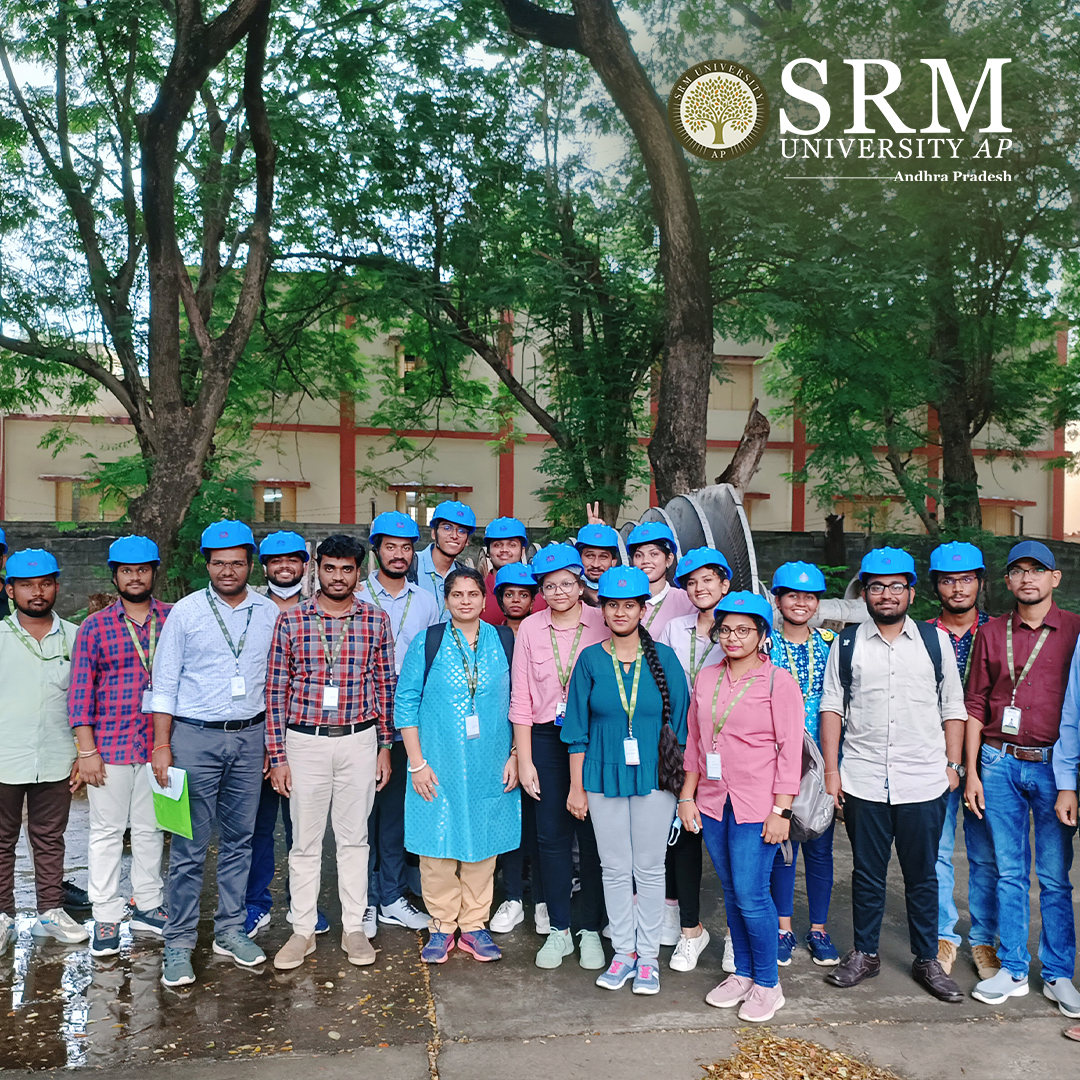 Industrial visit is an efficient learning strategy that nullifies the distance between academia and industry. Students and faculty of the Department of Electrical and Electronics Engineering visited Dr Narla Thatharao Thermal Power Station (NTTPS) on November 21, 2022. The objective of the visit was to give an overview of the Thermal Power plant, which is a coal-based power plant where coal is transported from coal mines to the power plant by railway in wagons or a merry-go-round system.
The state-of-the-art construction and technologies amazed the students as they experienced an insightful industrial visit that exposed them to the actual working of a thermal power plant while enforcing their theoretical knowledge of Power Systems, Control Systems and Electrical Machines. Students from semesters one to four participated in the industry visit. Dr Somesh Vinayak Tewari, Dr Shubh Lakshmi, Dr Bhamidi Lokeshgupta, Dr Ramanjaneya Reddy U, and Dr Venkata Ramireddy Y were the faculty who accompanied the students.
A Detailed Account of the Industry Visit
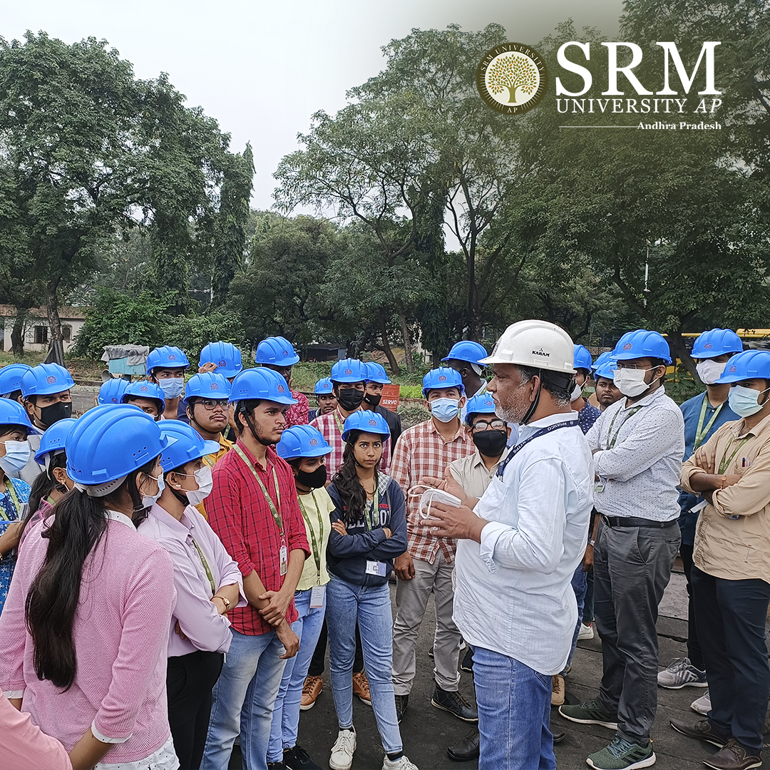 Coal is unloaded from the wagons using wagon tippler units to a moving underground conveyor belt. This coal from the mines is of no uniform size. So, it is taken to the Crusher house and crushed to a size of 20 mm. From the crusher house, the coal is either stored in dead storage, which serves as coal supply in case of coal supply bottleneck or to live storage in the raw coal bunker in the boiler house. Raw coal from the raw coal bunker is supplied to the Coal Mills by a Raw Coal Feeder. The Coal Mills or pulverizer pulverizes the coal. The powdered coal from the coal mills is carried to the boiler in coal pipes by high-pressure hot air. The pulverized coal air mixture is burnt in the boiler in the combustion zone.
Generally, in modern boilers, a tangential firing system is used where the coal nozzles/guns form a tangent to a circle. The temperature in the fireball is of the order of 1300˚C. The boiler is a water tube boiler hanging from the top. Water is converted to steam in the boiler, and steam is separated from water in the boiler Drum. The saturated steam from the boiler drum is taken to the Low-Temperature Superheater, Platen Superheater and Final Superheater, respectively, for superheating. The superheated steam from the final superheater is taken to the High-Pressure Steam Turbine (HPT). In the HPT, the steam pressure is utilised to rotate the turbine, and the resultant is rotational energy. From the HPT, the outcoming steam is taken to the Reheater in the boiler to increase its temperature as the steam becomes wet at the HPT outlet. After reheating, this steam is taken to the Intermediate Pressure Turbine (IPT) and then to the Low-Pressure Turbine (LPT). The outlet of the LPT is sent to the condenser for condensing back to water by a cooling water system. This condensed water is collected in the Hot well and sent to the boiler in a closed cycle. The rotational energy imparted to the turbine by high-pressure steam is converted to electrical energy in the Generator.
- Published in Departmental News, EEE NEWS, News
Dr Raviteja KVNS Received the Best Paper Award at TRACE 2022
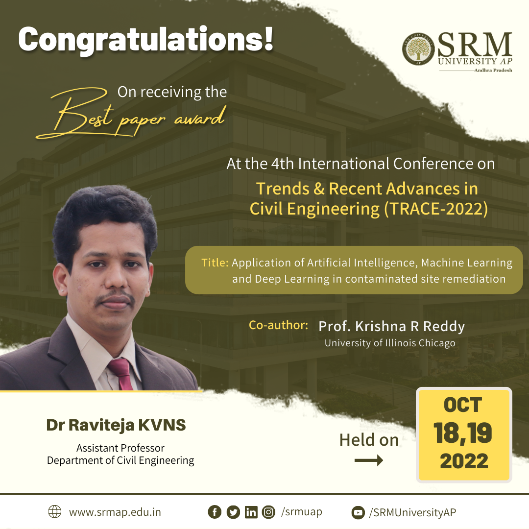 Soil and groundwater contamination is closely interlinked with human society because of its direct impact on population health and socioeconomic activities. The design and implementation of site remediation can be expensive, time-consuming, and may require much human effort. Emerging technologies such as Artificial Intelligence, Machine Learning, and Deep Learning have the potential to make site remediation cost-effective with reduced human effort.
Assistant Professor Dr Raviteja KVNS, Department of Civil Engineering, has received the Best Paper Award at the Fourth International Conference on Trends and Recent Advances in Civil Engineering (TRACE) 2022 for his paper Application of artificial intelligence, machine learning and deep learning in contaminated site remediation. The conference was held at Amity University, Uttar Pradesh, on October 18 and 19, 2022. His research reports the applications of AI and ML in contaminated site remediation.
Dr Raviteja’s future research plan includes studying potential applications of various AI, ML and DL techniques for Geotechnical and Geo-environmental design and testing applications so as to reduce the labours of physical and repetitive testing and associated human effort. This further improves precision as well as aids in decision-making. He has collaborated with Prof. Krishna R Reddy, University of Illinois Chicago, for this research work.
Abstract
Soil and groundwater contamination is caused by improper waste disposal practices and accidental spills, posing a threat to public health and the environment. It is imperative to assess and remediate these contaminated sites to protect public health and the environment as well as to assure sustainable development. Site remediation is inherently complex due to the many variables involved, such as contamination chemistry, fate and transport, geology, and hydrogeology. The selection of remediation method also depends on the contaminant type and distribution and subsurface soil and groundwater conditions. Depending on the type of remediation method, many systems and operating variables can affect the remedial efficiency. The design and implementation of site remediation can be expensive, time-consuming, and may require much human effort. Emerging technologies such as Artificial Intelligence, Machine Learning, and Deep Learning have the potential to make site remediation cost-effective with reduced human effort. This study provides a brief overview of these emerging technologies and presents case studies demonstrating how these technologies can help contaminated site remediation decisions.
- Published in CIVIL NEWS, Departmental News, Faculty Achievements, News, Research News

