All Management Events
- Dr Arpita Nayek June 30, 2023
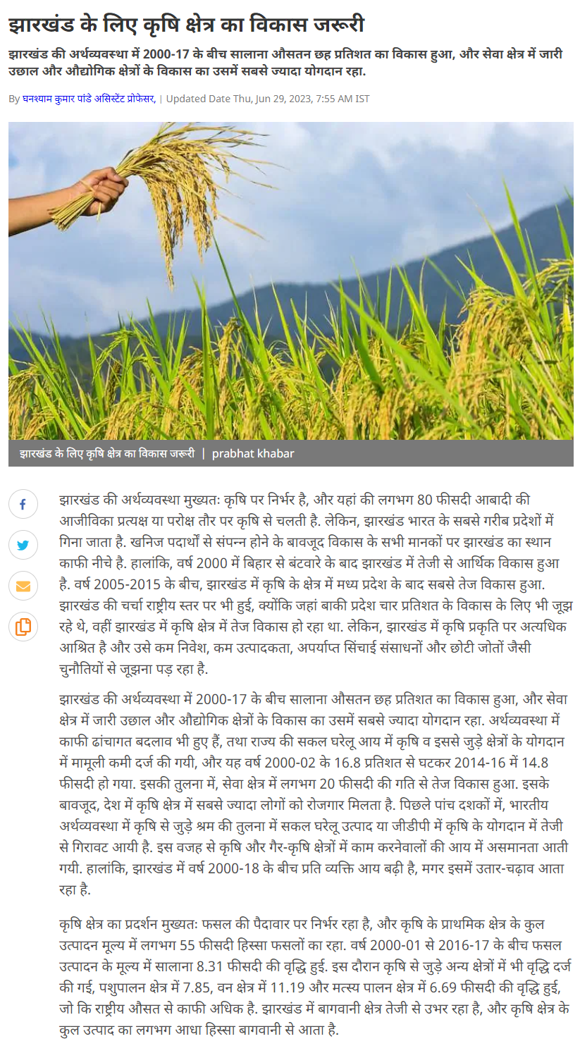
- Dynamics of agriculture development in Jharkhand after bifurcation June 30, 2023
DNP India
Continue reading →
DNP Education
Prabhat Kabar
- SRM University-AP Third in Nature Index Ranking June 30, 2023
Deccan Chronicle
Continue reading →
The Hans India
The Pioneer
Eenadu
Andhra Jyothi
Andhra Prabha
Andhra Patrika
Vartha Prapancham
Vartha
Visalaandhra
Surya
Power
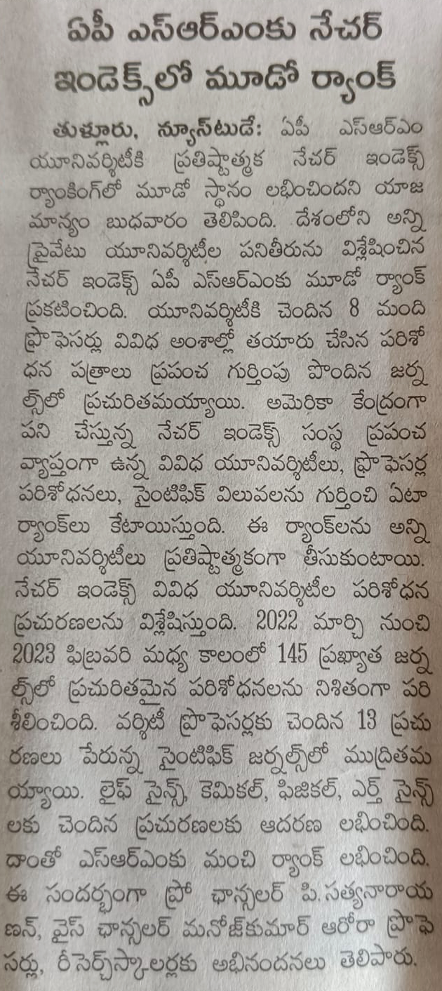
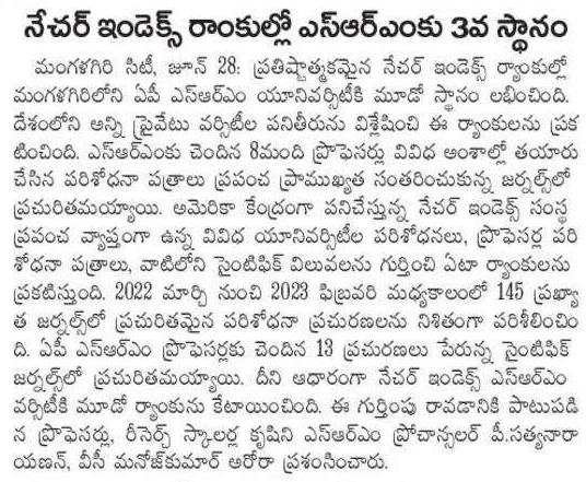
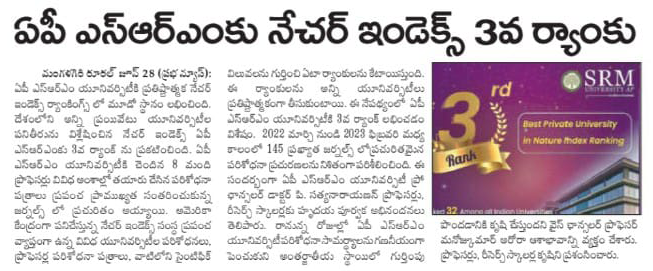
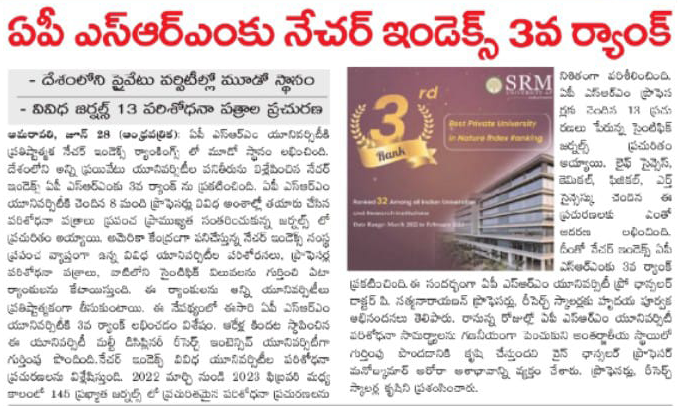
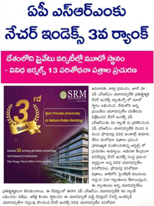
- India’s 3rd Best Private University in Nature Index Ranking for the Second Consecutive Time June 28, 2023
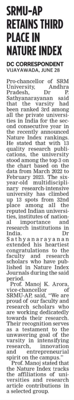
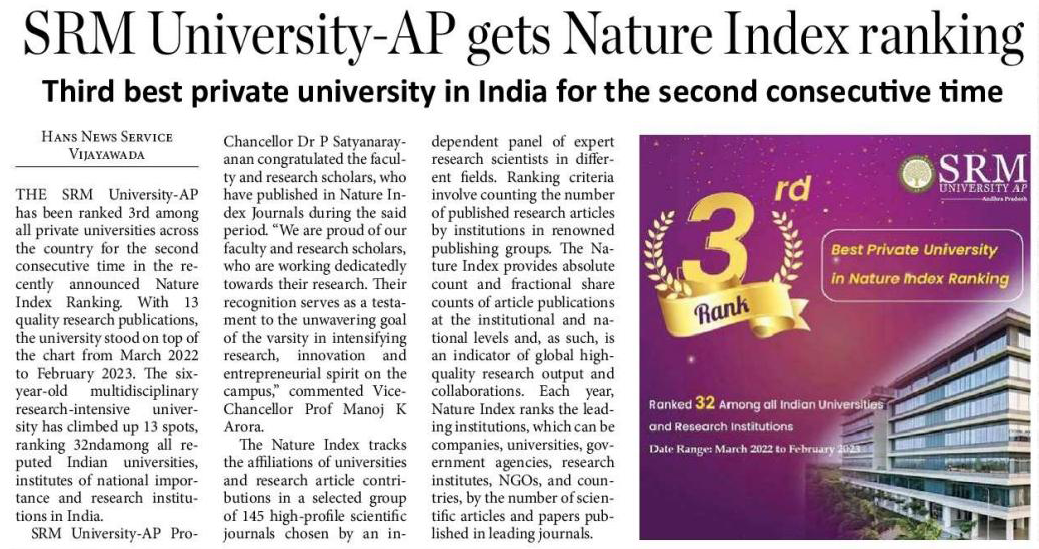
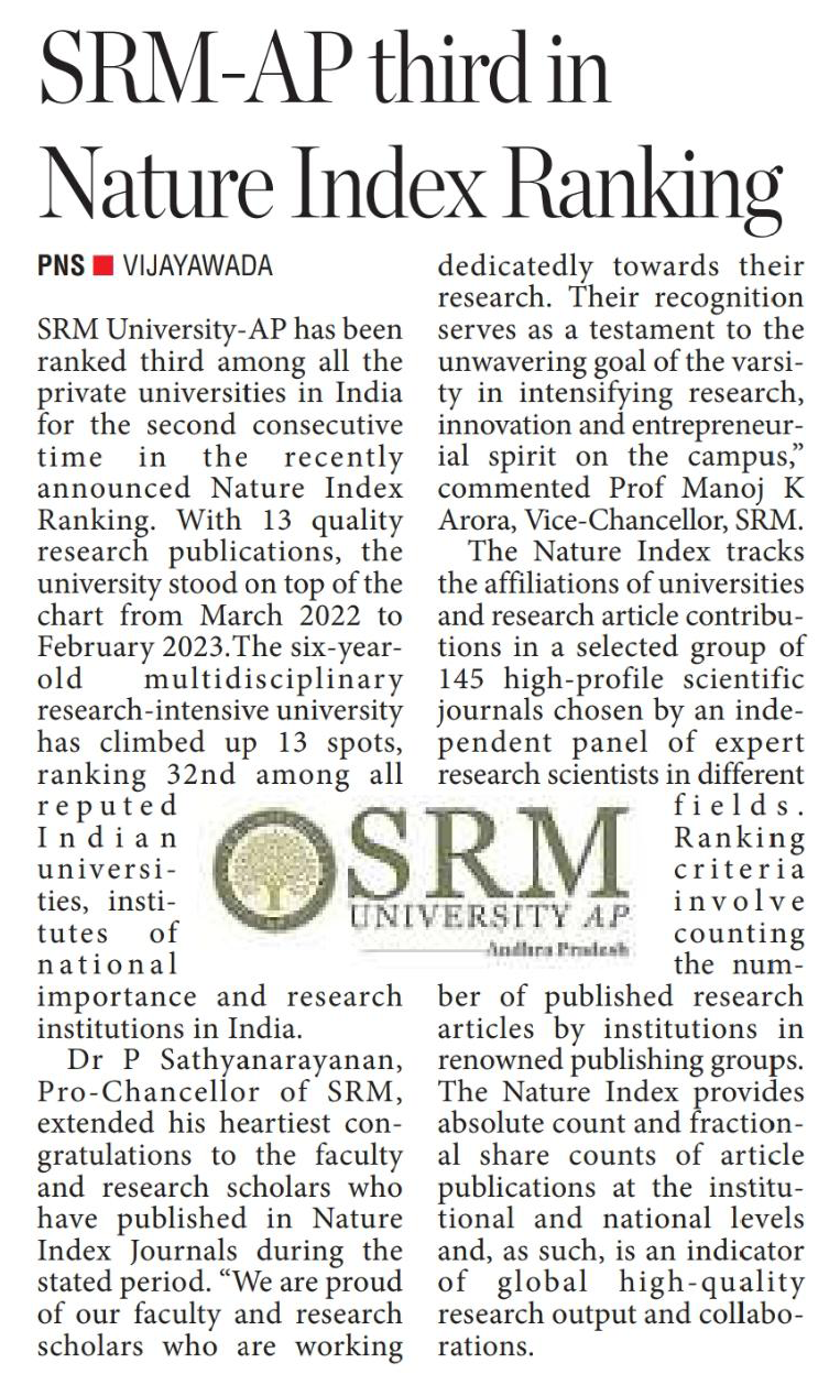
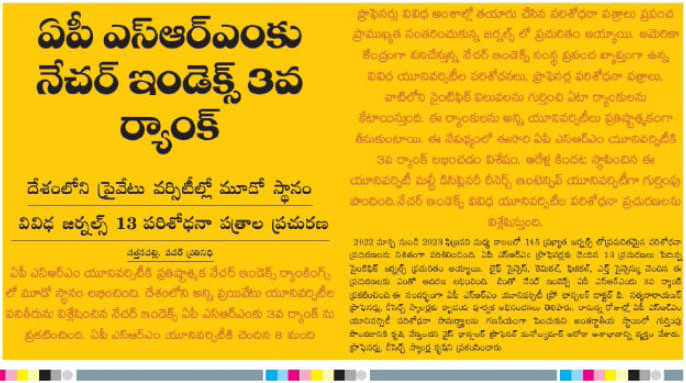
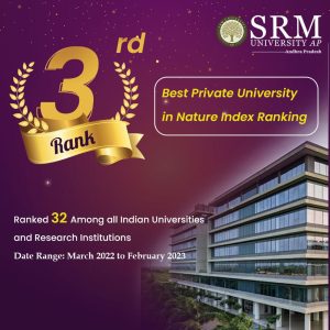 SRM University-AP has been ranked 3rd among all the private universities in India for the second consecutive time in the recently announced Nature Index Ranking. With 13 quality research publications, the university stood on top of the chart from March 2022 to February 2023. The six-year-old multidisciplinary research-intensive university has climbed up 13 spots, ranking 32nd among all reputed Indian universities, institutes of national importance and research institutions in India.
Dr P Sathyanarayanan, Pro-Chancellor of SRM University-AP, extended his heartiest congratulations to the faculty and research scholars who have published in Nature Index Journals during the stated period. “We are proud of our faculty and research scholars who are working dedicatedly towards their research. Their recognition serves as a testament to the unwavering goal of the varsity in intensifying research, innovation and entrepreneurial spirit on the campus’’, commented Prof. Manoj K Arora, Vice Chancellor of SRM University-AP.
The Nature Index tracks the affiliations of universities and research article contributions in a selected group of 145 high-profile scientific journals chosen by an independent panel of expert research scientists in different fields. Ranking criteria involve counting the number of published research articles by institutions in Renowned publishing groups. The Nature Index provides absolute Count and fractional Share counts of article publications at the institutional and national levels and, as such, is an indicator of global high-quality research output and collaborations.
Each year, Nature Index ranks the leading institutions, which can be companies, universities, government agencies, research institutes, NGOs, and countries, by the number of scientific articles and papers published in leading journals. The ranking involves research fields, such as life sciences, chemical sciences, physical sciences, and earth sciences. The Index helps assess research excellence by institutions, regions, and research disciplines.
Continue reading → - Dr Debabrata Senapati June 28, 2023
- World Environment Day Celebrations – An Initiative to Beat Plastic Pollution June 27, 2023
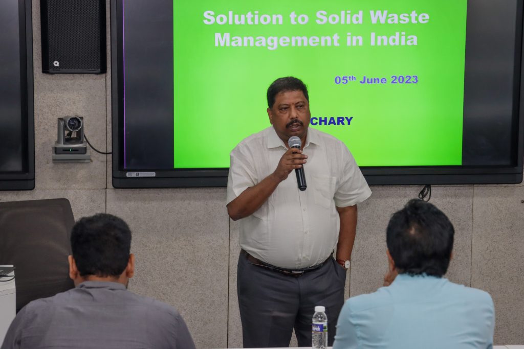
The Department of Environmental Science and Engineering celebrated World Environment Day on June 05, 2023, initiating awareness of the need to combat the climate crisis posed by global plastic pollution. The department organised two invited lectures from industry experts to expound on the theme of World Environment Day 2023, “Solutions to Plastic Pollution”. Efficient ways to tackle plastic pollution in the country and local waste management systems and policies were key points of discussion. Prof. K V Jayakumar, Professor, Department of Civil Engineering, National Institute of Technology, Warangal and Mr M V Chary, President, Jindal Urban Waste Management, Guntur, Andhra Pradesh, were the invited speakers.
Prof. K V Jayakumar lent thought-provoking insights into the types of water and how marketing strategies sell water at high prices to consumers. He also shared his experience of his visits to other countries in Europe and Asia where water is utilised wisely. Apart from water and plastic pollution, he also addressed the pressing issue of climate change happening over the years and the consequent increase in temperature. Prof. Jayakumar also suggested the book “Silent Spring” by Rachel Carson, which is often viewed as a landmark work of environmental writing, focusing on the harmful effects of pesticides on the environment.
Mr M V Chary shared in-depth information about the Jindal Urban Waste Management at Guntur, stating the various source of waste, collection points, transportation to the Jindal Waste – Energy Plant, and how waste is converted to energy (through incineration) at the plant. Unique aspects of the plant were also discussed at length, along with a video presentation demonstrating the complete working of the plant. The interactive session with Mr Chary, wherein he shared his expertise in the industrial sector, was beneficial for the students.
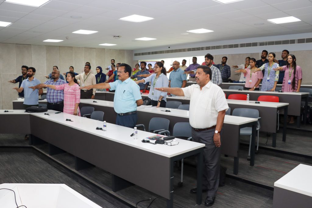
A pledge was taken by all participants at the end of the seminar emphasising the importance of saving the environment, sustainable resource management and the need of awareness among all in reducing the pollution as well as global warming and climate change.
Dr Karthik Rajendran, HOD of the Department, delivered the welcome address and the event witnessed an active participation of 85 participants which constitutes Professors, Faculty, PhD Scholars, Undergraduate students from various departments. There were also faculty and scholars from neighbouring colleges such as Acharya Nagarjuna University, Sidhartha Mahila Kalasala and Ministry of Culture, New Delhi.
Continue reading → - Dr Naresh Kumar Vemula June 26, 2023
- Physics Graduate Secures Two International Internships June 26, 2023
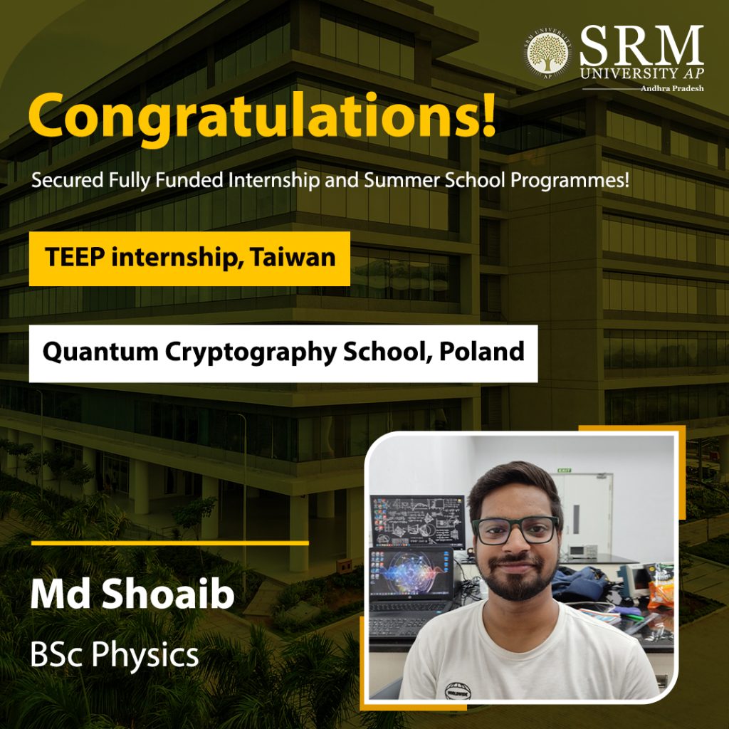
In an era where competition in the job market is on the peek, internships give you an edge over the rest of your peers. It gives you an opportunity to get real-time experience and put to practise what you have learnt. But not everyone manages to acquire an internship, while most students manage to barely secure a single internship, Md Shoaib, has bagged not one, but two exciting international internships in the field of Quantum Physics!
Here’s what Md Shoaib has to say on his remarkable achievement: “ I am excited to delve deeper into this topic and participate in enriching lectures, workshops, and interactions with experts in the field. It will be a valuable experience to learn from renowned Quantum scientists from various countries and broaden my understanding of Quantum Cryptography.”
Md Shoaib, a BSc Physics (Hons) with Research student has secured the prestigious Taiwan Experience Education Program (TEEP) internship programme along with a Summer School “Quantum Cryptography” internship offered by the University of Gdańsk, Poland. The internships not only offer a stipend but also covers travel, accommodation and logistic expenses.
We wish Md Shoaib the very best in his endeavour!
- Specialise in the Latest Tech with an MTech in Computer Science and Engineering June 26, 2023
With the advent of advanced technologies and innovations in the area of computer science and engineering, pursuing an MTech in Computer Science and Engineering has become a highly coveted career choice for students. An MTech degree in Computer Science and Engineering equips students with an extensive understanding of advanced computer concepts, including algorithms, machine learning, data structures, computer networks, and software engineering.
The MTech in Computer Science and Engineering curriculum is curated to provide individuals with hands-on experience in application development and computer programming, preparing them for a flourishing career in an array of industries, including artificial intelligence, software development, robotics, and cybersecurity. Furthermore, an MTech in Computer Science and Engineering also opens doors to endless opportunities for students to pursue research and development in leading-edge computer science disciplines.

What is MTech Computer Science Curriculum?
The MTech Computer Science and Engineering curriculum varies from institute to institute. However, most programmes incorporate a core set of courses that includes advanced topics in computer science and engineering. The MTech in Computer Science and Engineering syllabus typically incorporates a wide range of advanced subjects such as:
- Data Structures and Algorithms
- Operating Systems
- Computer Networks
- Software Engineering
- Artificial Intelligence
- Machine Learning
- Natural Language Processing
- Cybersecurity and Cryptography
- Distributed Systems
- Cloud Computing
- Robotics
These MTech in Computer Science subjects are curated to provide students with a rigid foundation in the area of computer science and engineering. In addition to the core courses, MTech Computer Science students also have the opportunity to specialise in a specific area of the domain through electives and research projects. These specialisations can include areas like robotics, distributed systems, cybersecurity, computer vision, and natural language processing.
Some MTech Computer Science curricula also require individuals to complete a research project or thesis, which enables them to use their knowledge and skills to solve a real-world problem in the domain.
What is MTech Computer Science Eligibility?
The MTech Computer Science eligibility depends on the institution offering the programme. Every institute has some general eligibility criteria for admission into its MTech Computer Science and Engineering programme. However, some common MTech Computer Science and Engineering eligibility criteria are as follows:
- The applicants must have completed a BTech degree in Computer Science and Engineering or a related discipline from a recognized institution.
- The applicant must have acquired a minimum of 50-60% aggregate marks in their undergraduate degree.
- Some institutes may also require applicants to have a valid GATE score in Computer Science and Engineering. Some institutes conduct their own entrance exam and require individuals to appear for them.
Stand Out in the Job Market with a World-class MTech in Computer Science and Engineering at SRM University-AP
SRM University-AP imparts top-notch MTech Computer Science and Engineering programme with a specialisation in emerging areas, including Artificial Intelligence & Machine Learning. The MTech Computer Science and Engineering curriculum at SRM AP is structured keeping in mind the need of the hour of the industry. The MTech graduates from SRM AP are eligible to either choose to work in the biggest giants of the industry or choose to pursue a PhD from a technical Institute of international repute. The courses of the programme are taught by highly qualified and skilled faculty who have overseas exposure to teaching and research.
MTech in Computer Science and Engineering specializations offered at SRM AP:
- Artificial Intelligence & Machine Learning
- Data Science
- Cyber Security
Moreover, the M.Tech. in Computer Science and Engineering eligibility at SRM AP requires students to have a basic degree or equivalent in BE / BTech in CSE / IT / SWE (or) MSc (CSE / IT) (or) MCA with GATE and a minimum of 60% aggregate marks.
To Conclude
MTech Computer Science and Engineering is ideal for individuals who want to enhance their technical skills and advance their careers. The programme assists them in specializing in an area of their choice, builds a strong foundation in advanced computer science and engineering topics, and offers promising opportunities to work with leading-edge technology. Pursuing an MTech degree in Computer Science and Engineering can provide you with a competitive edge in the job market and lead to enormous job roles and career growth.
Continue reading → - Shape the Future of Tech with BTech Computer Science and Engineering June 26, 2023
Living in the most populated country requires consistent innovation for betterment. Innovation requires innovative Computer Science Engineers. It is undeniable that Computer Science Engineers are highly in demand amid this tech-savvy era. Therefore becoming a Computer Science Engineer would be the wisest decision for a person. To become a Computer Science Engineer, one must pursue BTech in Computer Science and Engineering. Let us know first what BTech Computer Science and Engineering is.
BTech Computer Science and Engineering is an undergraduate course concentrating on developing skills in computer system technologies and programming terminology. The syllabus of BTech CSE is designed to provide comprehensive 21st-century skills like computer programming, computer application, research and development, analytics, data structure, etc. Let us discuss the BTech CSE syllabus in detail.

BTech Computer Science and Engineering is one of the most demanded courses in the country as the BTech CSE subjects taught during the course impart and enhance industry-relevant skills among students so that they become highly knowledgeable, skilled graduates who can work anywhere in the world. Some of the major subjects taught during BTech CSE are:
- Software Engineering
- Artificial Intelligence
- Computers and Information Technology
- Database Information System
- Applied Physics
Any course is lucrative only when its scope is wide and prosperous. BTech in Computer Science and Engineering is a popular course as its scope is wide and magnificent in monetary terms. As per the reports, the salary package of a BTech Computer Science Engineer is much higher than any other graduate courses. Some of the best career options available for computer science engineers are:
- Software Engineer
- Data Engineer
- Software Architect
- E-commerce Business Analyst
- Full-stack Developer
- Computer Hardware Engineer
- Network Engineer
- Computer Scientist
By now, it has been established that BTech Computer Science and Engineering is a widely popular course among young aspirants. The benefits of being a computer science engineer are quite prosperous when they have a name of a good university with them. Pursuing a creditable course is important, but pursuing that course from a renowned university is equally important to get celebrated perks. One of the most renowned universities, especially for providing world-class education, is SRM University-AP.
Step Into the Digital World with BTech in Computer Science and Engineering at SRM University-AP
SRM University-AP, falls under the top-ranking universities in India. The University aims to create and impart knowledge and bring forth a unique learning experience to its students who become technologically advanced and efficient resources to society. The Department of Computer Science and Engineering at SRM AP strives to contribute to this tech revolution by transforming its students into savvy professionals with firm know-how of state-of-the-art software and hardware mechanisms.
BTech in Computer Science and Engineering is the University’s most coveted programme as the Department of CSE provides research-based education and follows an Active-learning pedagogy to improve the quality of the learning experience for the students. The University has installed state-of-the-art laboratories to encourage students to carry out projects of multidisciplinary nature. It gives students opportunities to get international exposure in reputed foreign institutes like Northeastern University, Flinder University, the University of Wisconsin-Madison, Asia University etc.
BTech CSE SRM University-AP covers various computer software and hardware topics and is taught by highly experienced faculty members. Some specialisations offered under the course are:
- Artificial Intelligence and Machine Learning
- Internet of Things (IoT)
- Big Data Analytics
- Cyber Security
- Distributed and Cloud Computing
Overall, BTech CSE students at SRM AP are given high-quality research-oriented education that will shape them to succeed in the modern, developing technological world.
Be Future-ready with BTech CSE
BTech Computer Science and Engineering is dandy among young scholars. It provides all the 21st-century technological skills that make students efficient and skilful who are dexterous and capable of working for newly developed and developing organisations. The scope of BTech CSE is wide and offers graduates a safe and secure future.
SRM University-AP aims to provide world-class education to its students by providing highly-qualified faculty and a world-class curriculum. The University thrives in helping its graduates through the teaching-learning process at the time of placement and even after that so that the students always get constant support from the University. So what are you waiting for? Enrol in the BTech CSE programme at SRM AP and secure your future with constant growth.
Continue reading →