School of Engineering and Sciences
Department of Electronics and Communication Engineering


Breast cancer (BC) is one of the most common types of cancer among women with a high mortality rate. Histopathological analysis facilitates the detection and diagnosis of BC but is a highly time-consuming specialised task, dependent on the experience of the pathologists. Hence, there is a dire need for computer-assisted diagnosis (CAD) to relieve the workload on pathologists. Dr Sudhakar Tummala, Assistant Professor, Department of Electronics and Communication Engineering, has conducted breakthrough research on this domain in his paper titled BreaST-Net: Multi-Class Classification of Breast Cancer from Histopathological Images Using Ensemble of Swin Transformers published in the Q1 Journal Mathematics, having an Impact Factor of 2.6.
Abstract
Breast cancer (BC) is one of the deadly forms of cancer and a major cause of female mortality worldwide. The standard imaging procedures for screening BC involve mammography and ultrasonography. However, these imaging procedures cannot differentiate subtypes of benign and malignant cancers. Therefore, histopathology images could provide better sensitivity toward benign and malignant cancer subtypes. Recently, vision transformers are gaining attention in medical imaging due to their success in various computer vision tasks. Swin transformer (SwinT) is a variant of vision transformer that works on the concept of non-overlapping shifted windows and is a proven method for various vision detection tasks. Hence, in this study, we have investigated the ability of an ensemble of SwinTs for the 2- class classification of benign vs. malignant and 8-class classification of four benign and four malignant subtypes, using an openly available BreaKHis dataset containing 7909 histopathology images acquired at different zoom factors of 40×, 100×, 200× and 400×. The ensemble of SwinTs (including tiny, small, base, and large) demonstrated an average test accuracy of 96.0% for the 8-class and 99.6% for the 2-class classification, outperforming all the previous works. Hence, an ensemble of SwinTs could identify BC subtypes using histopathological images and may lead to pathologist relief.
A brief summary of the research in layperson’s terms
Breast cancer (BC) is the second deadliest cancer after lung cancer, causing morbidity and mortality worldwide in the women population. Its incidence may increase by more than 50% by the year 2030 in the United States. The non-invasive diagnostic procedures for BC involve a physical examination and imaging techniques such as mammography, ultrasonography and magnetic resonance imaging. However, the physical examination may not detect it early, and Imaging procedures offer low sensitivity for a more comprehensive assessment of cancerous regions and identification of cancer subtypes. Histopathological imaging via breast biopsy, even though minimally invasive, may provide accurate identification of the cancer subtype and precise localization of the lesion. However, this manual examination by the pathologist could be tiresome and prone to errors. Therefore, automated methods for BC subtype classification are warranted.
Deep learning has revolutionised many areas in the last decade, including healthcare for various tasks such as accurate disease diagnosis, prognosis, and robotic-assisted surgery. There were studies based on deep convolutional neural networks (CNN) for detecting BC using the aforementioned imaging procedures. However, CNNs exhibit inherent inductive bias and are variant to translation, rotation, and location of the object of interest in the image. Therefore, image augmentation is generally applied while training CNN models, although the data augmentation may not provide expected variations in the training set. Hence, self-attention based deep learning models that are more robust towards the orientation and location of an object of interest in the image are rapidly growing.
SwinTs are an improved version of earlier vision transformer (ViT) architecture and are hierarchical vision transformers using shifted windows that work based on self-attention. For efficient modelling, self-attention within local windows was proposed and computed, and to evenly partition the image, the windows are arranged in a non-overlapping manner. The window-based self-attention has linear complexity and is scalable. However, the modelling power of window-based self-attention is limited because it lacks connections across windows. Therefore, a shifted window partitioning approach that alternates between the partitioning configurations in consecutive Swin transformer blocks was proposed to allow cross-window connections while maintaining the efficient computation of non-overlapping windows. The shifted window scheme in Swin transformers offers increased efficiency by restricting self- attention computation to local windows that are non-overlapping while also facilitating a cross-window connection. Overall, the SwinT network’s performance was superior to that of the standard ViTs.
Therefore, the paper analyses the ability of an ensemble of Swin transformer models (BreaST-Net) for the automated multi-class classification of BC by investigating histopathological images. The work dealt with both benign and malignant subtypes. Further, the benign cancer subtypes include fibroadenoma, tubular adenoma, phyllodes tumour, and adenosis. Whereas the malignant subtypes contain ductal carcinoma, papillary carcinoma, lobular carcinoma, and mucinous carcinoma.
Social implications of the research
Dr Sudhaker Tummala explains that the computer-aided subtyping of breast cancer from histopathology images using an ensemble of fine-tuned SwinT models can be an alternative to manual diagnoses, thereby reducing the burden on clinical pathologists.
Collaborations
In the future, Dr Tummala will advance his research to add explainability to the ensemble model predictions and also to develop models that can work on fewer data samples.
Continue reading →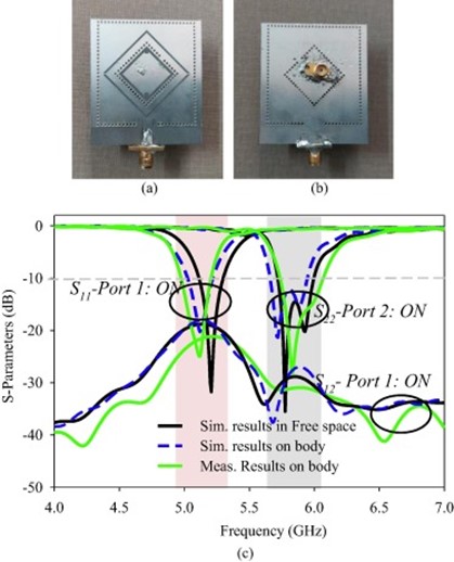
With the recent advancements in modern wireless body area network (WBAN) communication, the demand for compact low-profile wireless computing devices has witnessed a vast increase. Consequently, the antennas which play a critical role in this network are developed with different polarization in distinct frequency bands so as to maintain better reliability of communication links. Dr Divya Chaturvedi, Assistant Professor, Department of Electronics and Communication Engineering, has published a paper titled, “A Dual-Band Dual-Polarized SIW Cavity-Backed Antenna-Duplexer for Off-body Communication” as first author in the Q1 Journal AEJ – Alexandria Engineering Journal having an impact factor of 6.77. The paper discusses the self-duplexing antennas, offering two channels for concurrent transmission and reception, leading to a simple and compact transceiver.
Abstract
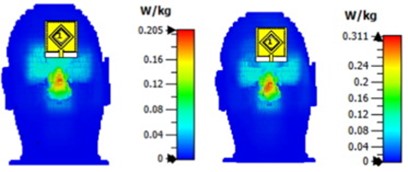
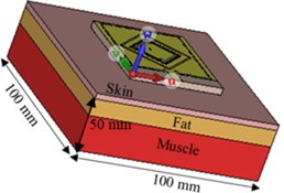
A novel dual-band, dual-polarized antenna-duplexer scheme is intended to be used for WLAN 802.11a and ISM band applications using Substrate Integrated Waveguide (SIW) Technology. The antenna consists of two planar SIW cavities of different dimensions where a smaller sized diamond- shaped cavity is inserted inside the larger rectangular cavity to share the common aperture area. The diamond-ring shaped slots are etched in each cavity for radiation. The larger diamond ring slot is excited with a microstrip feedline to operate at 5.2 GHz while the smaller slot is excited with a coaxial probe to operate at 5.8 GHz. The antenna produces linear polarization at 5.2 GHz (5.1–5.3 GHz) due to the merging of TE 110 and TE 120 cavity modes while circular polarization around 5.8 GHz due to orthogonally excited TM100 and TM010 modes (5.68–5.95 GHz). The slots are excited in an orthogonal fashion to maintain a better decoupling between the ports (i.e. –23 dB). The performance of the antenna has been verified in free space as well as in the vicinity of the human body. The antenna offers the gain of 6.2 dBi /6.6 dBi in free space and 5.8 dBi / 6.4 dBi on-body at lower-/ higher frequency-bands, respectively. Also, the specific absorption rate (SAR) is obtained < 0.245 W/Kg for 0.5 W input power averaged over 10 mW/g mass of the tissue. The proposed design is a low-profile, compact single-layered design, which is a suitable option for off-body communication.
Explanation of the research in layperson’s terms
The paper further expounds on the social implication of this innovative research. Dr Chaturvedi explains that the antenna, being dual-band and dual-polarized, can function as a transceiver circuit. Due to different polarization, it can operate in both the frequency bands simultaneously without affecting the performance. In the first frequency band at 5.2 GHz, it can link with Wi-Fi and in the second frequency band at 5.8 GHz, it is able to communicate with antennas placed in other medical instruments which are used in the vicinity of the human body.
Collaborations
1. Dr Arvind Kumar, Assis. Professor, b Department of Electronics and Communication
Engineering, VNIT Nagpur, India
2. Dr Ayman A Althuwayb, Department of Electrical Engineering, College of Engineering,
Jouf University, Sakaka, Aljouf 72388, Saudi Arabia

Primary brain tumours make up less than 2% of cancers and statistically occur in around 250,000 people a year globally. Medical resonance imaging (MRI) plays a pivotal role in the diagnosis of brain tumours and advanced imaging techniques can precisely detect brain tumours. On this note, Dr Sudhakar Tummala, Assistant Professor, Department of Electronics and Computer Engineering, has published a paper titled, “Classification of Brain Tumour from Magnetic Resonance Imaging using Vision Transformers Ensembling” in the journal Current Oncology having an impact factor of 3.1. The paper highlights the pioneering breakthrough made in the development of vision transformers (ViT) in enhancing MRI for efficient classification of brain tumours, thus reducing the burden on radiologists.
Abstract of the paper
The automated classification of brain tumours plays an important role in supporting radiologists in decision making. Recently, vision transformer (ViT)-based deep neural network architectures have gained attention in the computer vision research domain owing to the tremendous success of transformer models in natural language processing. Hence, in this study, the ability of an ensemble of standard ViT models for the diagnosis of brain tumours from T1-weighted (T1w) magnetic resonance imaging (MRI) is investigated. Pretrained and fine tuned ViT models (B/16, B/32, L/16, and L/32) on ImageNet were adopted for the classification task. A brain tumour dataset from figshare, consisting of 3064 T1w contrast-enhanced (CE) MRI slices with meningiomas, gliomas, and pituitary tumours, was used for the cross-validation and testing of the ensemble ViT model’s ability to perform a three-class classification task. The best individual model was L/32, with an overall test accuracy of 98.2% at 384 × 384 resolution. The ensemble of all four ViT models demonstrated an overall testing accuracy of 98.7% at the same resolution, outperforming individual model’s ability at both resolutions and their ensemble at 224 × 224 resolution. In conclusion, an ensemble of ViT models could be deployed for the computer-aided diagnosis of brain tumours based on T1w CE MRI, leading to radiologist relief.
A brief summary of the research in layperson’s terms
Brain tumours (BTs) are characterised by the abnormal growth of neural and glial cells. BTs causes several medical conditions, including the loss of sensation, hearing and vision problems, headaches, nausea, and seizures. There exist several types of brain tumours, and the most prevalent cases include meningiomas (originate from the membrane surrounding the brain), which are non-cancerous; gliomas (start from glial cells and the spinal cord); and glioblastomas (grow from the brain), which are cancerous. Sometimes, cancer can spread from other parts of the body, which is called brain metastasis. A pituitary tumour is another type of brain tumour that develops in the pituitary gland in the brain, and this gland primarily regulates other glands in the body. Magnetic resonance imaging (MRI) is a versatile imaging method that enables one to noninvasively visualise inside the body, and is in extensive use in the field of neuroimaging.
There exist several structural MRI protocols to visualise inside the brain, but the prime modalities include T1-weighted (T1w), T2-weighted, and T1w contrast-enhanced (CE) MRI. BTs appear with altered pixel intensity contrasts in structural MRI images compared with neighbouring normal tissues, enabling clinical radiologists to diagnose them. Several previous studies have attempted to automatically classify brain tumours using MRI images, starting with traditional machine learning classifiers, such as support vector machines (SVMs), k-nearest-neighbour (kNN), and Random Forest, from hand-crafted features of MRI slices. With the rise of convolutional neural network (CNN) deep learning model architectures since 2012, in addition to emerging advanced computational resources, such as GPUs and TPUs, during the past decade, several methods have been proposed for the classification of brain tumours based on the finetuning of the existing state-of-the-art CNN models, such as AlexNet, VGG16, ResNets, Inception, DenseNets, and Xception, which had already been found to be successful for various computer vision tasks.
Despite the tremendous success of CNNs, they generally have inductive biases, i.e., the translation equivariance of the local receptive field. Due to these inductive biases, CNN models have issues when learning long-range information; moreover, data augmentation is generally required for CNNs to improve their performance due to their dependency on local pixel variations during learning.Therefore, in this work, the ability of pretrained and fine tuned ViT models, both individually and in an ensemble manner, is evaluated for the classification of meningiomas, gliomas, and pituitary tumours from T1w CE MRI at both 224 × 224 and 384 × 384 image resolutions.
Dr Sudhakar Tummala has mentioned the social implications of the research by expounding that the computer-aided diagnosis of brain tumours from T1w CE MRI using an ensemble of fine tuned ViT models can be an alternative to manual diagnoses, thereby reducing the burden on clinical radiologists. He also explains the future prospects of his research, which is to add explainability to the ensemble model predictions and to develop methods for precise contouring of tumour boundaries.
Details of Collaborations
Prof Seifedine Kadry, Department of Applied Data Science, Noroff University College, Kristiansand, Norway.
Dr Syed Ahmad Chan Bukhari, Division of Computer Science, Mathematics and Science, Collins College of Professional Studies, St. John’s University, New York, USA.
Continue reading →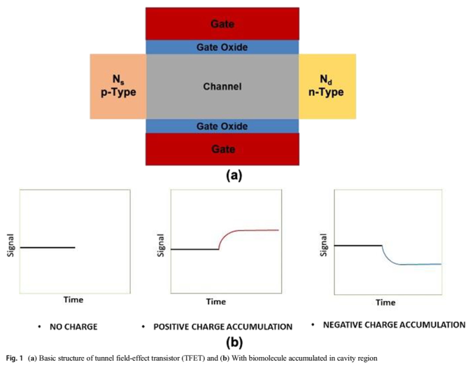
The Department of Electronics and Communication Engineering is glad to announce that Mr Garikapati Anith Chowdary, a BTech passed-out student has published a paper in collaboration with Assistant Professor Dr M Durga Prakash. The paper titled Tunnel Field Effect Transistor Design and Analysis for Biosensing Applications was published in the Q2 journal Silicon having an Impact Factor 2.941.
The physical modelling of the tunnel field effect transistor (TFET) is done in this study. The Silvaco TCAD tool is used to design and simulate the TFET structure. The FET device has attracted a lot of attention as the ideal tool for creating biosensors because of its appealing properties such as ultra-sensitivity, selectivity, low cost, and real-time detection capabilities in a sensing point of view.
These devices have a lot of potential as a platform for detecting biomolecules. Short channel effects, specificity, and nano-cavity filling have all been improved in FET-based biosensors. FET-based biosensors are appropriate for label-free applications. Random dopant variations and a thermal budget are seen during the construction of a JLFET. To overcome this problem, the charge-plasma-based concept was established in FETs in this study.
Different metallurgical functions for electrodes were employed in this biosensor to behave as a p-type source and n-type drain. To alleviate the short channel effects, a dual material gate work function for the gate electrode was devised, as well as a double gate architecture. Biomolecules can be neutral or charge-based, and both types of biomolecules can be identified using a proof-of-concept FET-based biosensor. Changes in the drain current (Id) of the device were achieved by varying dielectric values and charges in the cavity region with variable cavity lengths.
Continue reading → Research at the Department of Electronics and Communication Engineering is currently developing defect detection algorithms. Assistant Professor Dr V Udaya Sankar, Professor Dr Yellampalli Siva Sankar, and their BTech student Ms Gayathri Lakshmi have published a paper, A Review of various defects in PCB, in the Journal of Electronic Testing: Theory and Applications with an impact factor of 0.795.
Abstract
Printed Circuit Boards (PCBs) are the building blocks for all electronic products. Fabrication of a PCB involves various mechanical and chemical processes. As obtaining accuracy in the mechanical and chemical processes is very difficult, various defects/faults are formed during PCBs fabrication. These fabrication defects lead to performance degradation of electronic products. This paper describes multiple defects present in PCBs under the Through-hole and SMD categories. To understand the frequency of occurrence and reason for defects in both manual and machine, PCB fabrication data was collected and analysed from April 2017 to July 2020 as a part of industry collaboration.
The research is a review done on the defects present in PCB. Researchers surveyed various papers on PCB defects and their detection. Based on the literature review and information obtained from Efftronics systems Pvt. Ltd, they classified the defects, gave a detailed explanation for each, and provided some analysis of their occurrences.
While doing the literature review, researchers observed that no paper mentioned all the defects that can occur in the case of PCB fabrication. For this reason, they came up with this paper which provides detailed information regarding the defects. Information is also obtained from the industry. Comparing the defects can help focus on the critical defects for future research on defect detection methodology.
The project is done in collaboration with Efftronics Systems Pvt. Ltd. Through the partnership, the company supported sharing images, insights information related to defects and involved in discussions. Also, the company allowed visiting their premises to understand more about PCB defects. Researchers look forward to creating a prototype that detects all the defects mentioned in this paper for a given PCB.
Continue reading →